Tuesday, March 18, 2014
Friday, March 14, 2014
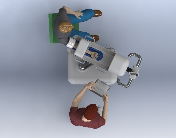
Robot to draw blood automatically
Fantastic development: a startup company (check them out at: www.veebot.com) has developed a robot that draws blood. The whole process takes a minute and is fully automated. It finds the right vein and draws the blood automatically.
Check out this coverage at IEEE Spectrum Profile: Veebot
Check out this coverage at IEEE Spectrum Profile: Veebot
Monday, March 10, 2014
ECR 2014 - RC1605 - Improving workflow efficiency and quality 2
reporting has four strategies:
1. text mode
2. fill in the blanks
3. serial assembly
4. mixed or nested assembly
Many different factors have been published that relate to errors in radiology reporting. Therefore, a more structured approach to reporting is required since the images are not the product of radiology but the conclusion of the report is.
The ESR and RSNA has presented a joint initiative on structured reporting. The different affiliated, subspecialty and national societies of the ESR will be uploading structured reports through the website of the RSNA. The IHE MRRT Management of Radiology Report Templates defines the technical framework of building report templates.
The HTML reports downloadable from the RSNA repository can be used and locally adapted to have optimal reports.
In the coming years ESR subcommittees should start to contribute to the RSNA and although practical clinical impelentations are limited, interest from radiologists and willingness to start testing these report is growing.
ECR 2014 - RC1605 - Improving workflow efficiency and quality 1
decision support in radiology is required because of the large variety of examinations. For example, double scanning rates can go up to 30% and many unnecessary exams are conducted. The decision of the appropriate imaging is heavily depending on the availability of the clinical inforrmation to properly use scoring lists to determine the optimal imaging.
The challenge with implementation of computerized physician order entry is the availability of proper rules and guidelines.
The ACR has developed an IT solution to provide decision support in the US with a commercial product called ACRSelect that can be used as a stand-alone, web based, or EPR integrated solution.
In Europe decision support is hampered by te variety in countries and languages. Only the UK and France have guidelines and recommendations but because of the wide variety between the UK and France guidelines, the ACR approach is adopted. There is interest to develop a set of global guidelines together with ACR.
CDS could help improve medical quality and effiiciency, avoid unnecessary imaging thus reducing cost and reducing radiation exposure.
Friday, March 7, 2014
ECR 2014 - SF7b - Imaging biomarkers in cancer drug development
An area where medical imaging informatics could play a major role is imaging biomarkers. The involvement of imaging informatics in this area is not only in the image processing, but also in validation, evaluation and collection of imaging data and related quantitative data using imaging biobanks.
Imaging biomarkers are currently a hot topic, in imaging this relates to:
1. size measurement based imaging biomarkers (RECIST, mRECIST and EASL)
2. functional and molecular biomarkers (perfusion, vascular function, cellularity, cell-death, metabolism, proliferation, and target specific tracers)
there are three biomarker modalities:
1. biospecimen removed from a patient
2. biosignal measured using in vivo device
3. patient reported outcomes
Important is that biomarkers require extensive validation and standardization of acquisition, evaluation, and measurement.
The validation of imaging biomarkers however, is a very hard job.
Imaging biomarkers should be used to better identify the patients most likely to benefit and to obtain better hypothosis testing.
problems in using imaging biomarkers include the variety in accquisition devices and the fact they are not designed for biomarker measurements.
Thursday, March 6, 2014
ECR 2014 - RC305 - Mobile IT in radiology
In radiology, display usages is usually in portrait and sometimes in landscape orientation. The introduction of tablets has introduced the ability to mix the use of portrait and landscape use of a display. The tablet also provides many more features such as speech recognition that are normally provided by the PACS workstation. Tablets allow portability of images outside radiology, facilitates interaction wiith clinicians, facilitates interaction with patients.
In this session a good overview was given by J. Fernandez-Bayo about the technological developments showing a close to diagostic trend in the capabilities of tablets. After this, O. Ratib presented on the software side of tablets and the possible applications in Radiology. Good applications are already shown and validated although clinical question at a time. The big breakthrough now is the use in surgery where iPads are brought into the OR to have access to the images of a patient. Currently the images are downloaded to the iPad but the trend is towards cloud computing with on-line acces to data and high end visualization and post-processing.
In a final presentation, E. Randschaert presented on teleradiology. In a very interesting presentation he showed that mobile devices are not yet to be used for routine primary diagnosis, but they are moving in that way. The developments already allow a wide spread implementation and the tables are already used by many physicians for different applications ranging from image viewing to teaching.
ECR 2014 - session SS105 - Trends in reporting, image management and mobile computing
10 presentations on different topics were covered in this scientific session:
CAD
Evaluation of a Siemens software tool called FastSpine to perform analysis of the spine with automatic labeling and curved reconstruction. They showed a high percentage of correct automatic labeling of 93.5%. FastSpine took about a minute to perform on average with manual correction where needed. The CAD provided time saving in more complex cases and had a comparable performance.
Two presentations focused on RECIST implementation. One looked at the implementation of dedicated tools for RECIST analysis and showed a clear saving in reading time while maintaing reproducibility. A Japanese group presented a cloud-based Lesion Management Solution that is used to track patients in lung cancer screening workup.
MOBILE DEVICES
This session showed again that mobile devices are of great interest and interesting applications are identified and implemented. Four of the presentations covered this topic.
A by dr von Falk showed that an iPad could be used for diagosis of CT data although screen size and security are still hampering the implementation. Another issue with mobile devices is the diiffering reading environments that cannot be controlled and thus could influence the reading quality. Mobile devices should probably be implemented as additional possibility and not for primary use. The dkfz demonstrated the use of an iPad for offline analysis of radiology information at a distant location to be used during autopsy.
A group from Pisa also evaluated the iPad on CT images and showed the iPad to be faster for evaluation with even higher sensitivity and specificity for the iPad.
An important point to keep in mind with all the evidence for using iPads in radiology is that it is all focused on a limited, well defined, clinical question and mostly limits to CT.
Another group from Germany introduced the iPad in anatomical courses. The developed AnatomyMap a dedicated software tool for interactive exploration of volume rendered data running on a remote server using a web based interface.
QUALITY CONTROL
A group from switzerland showed that after transfer from free text to structured reporting they needed quaity control of those structured reports. They analysed about 250 reports on over 30 criteria. They showed that one third was excellent, one third good and one third non-optimal. The two worst performing radiologists were those not (properly) using the structured reporting.
USABILITY
A Dutch group presented their work on interface customization. Most PACS systems have an adaptable interface, but the radiologist do not optimally use this capability. The showed that using adaptive support suggestions based on usage could help to improve radiology workflow and workstation usage.
Subscribe to:
Posts (Atom)